

Vascular Ultrasound: Checking Circulation and Blood Flow
Cayde Omura, RVT Vascular Sonographer

A vascular ultrasound is a non-invasive imaging test that uses sound waves to evaluate the health and function of your blood vessels. This simple, painless test plays a vital role in detecting circulation problems before they lead to serious health concerns.
At Hawaii Heart Associates, we use vascular ultrasound to examine different parts of the body depending on your symptoms and risk factors:
Aortic Ultrasound: Checks for aneurysms or enlargement of the abdominal aorta. Detecting these early can help prevent life-threatening complications.
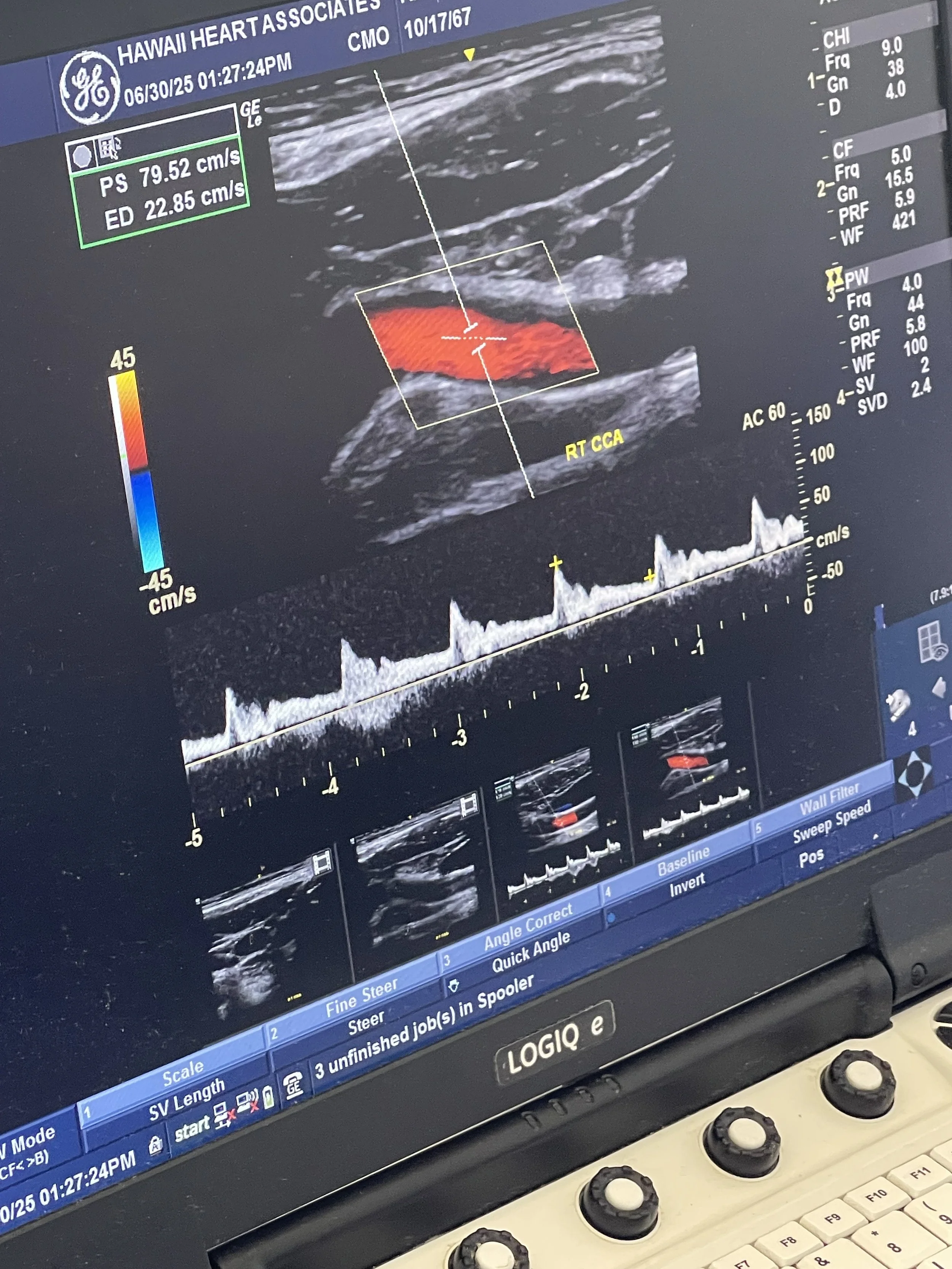
Carotid Ultrasound: Evaluates the carotid arteries in the neck for narrowing or blockages, which can increase your risk of stroke.
Lower Extremity Venous Ultrasound: Assesses the veins in the legs for blood clots (deep vein thrombosis) or signs of venous insufficiency, which may cause leg pain, swelling, or varicose veins.
Lower Extremity Arterial Ultrasound: Measures blood flow in the leg arteries and helps detect peripheral artery disease (PAD)—a condition that can cause pain, ulcers, or, if left untreated, even limb loss.
These tests are performed using a handheld device called a transducer, which glides over your skin after a small amount of gel is applied. You’ll be awake, comfortable, and usually done in less than an hour.
Why it matters:
Vascular ultrasounds are essential for early detection, risk assessment, and guiding treatment—helping to prevent strokes, aneurysms, or other serious vascular complications..